Skip the X-Ray, Get a CT Scan
Conventional dental implants may not be a suitable tooth replacement solution for everyone for a variety of medical reasons. One of the primary reasons a patient seeking traditional dental implants might not be a candidate is due to a lack of adequate jaw bone depth.
A CT scan of the oral cavity, particularly on the site of the prospective dental implantation, before an implant procedure is critical. It can help assess a patient’s suitability for an implant procedure, including detailed visualization of existing underlying jaw bone depth that can help determine whether or not a patient may require a bone graft.
A diagnostic CT scan is also vital for the accurate, precise, and safe placement of the implant itself with as few complications as possible. Essentially, a CT scan before a dental implant procedure will significantly improve the likelihood of a seamless, successful procedure and a smooth recovery thereafter.

ArtLab Dentistry Has Our Own Revolutionary CT Scanner
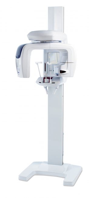
ArtLab Dentistry has our own revolutionary hi-tech CT Scanner which represents a new frontier in x-ray diagnostics. It is the latest 3D high-resolution J Morita 3D R100 scanner, which has the longest history and the lowest dosage of radiation on the market. We can obtain the most accurate possible view of a patient’s mouth needed for treatment. This also means we can instantly design and plan your implants with full control. We do not have to waste precious time waiting for a third party to provide your scan.
ArtLab Dentistry has our own revolutionary hi-tech CT Scanner which represents a new frontier in x-ray diagnostics. It is the latest 3D high-resolution J Morita 3D R100 scanner, which has the longest history and the lowest dosage of radiation on the market. We can obtain the most accurate possible view of a patient’s mouth needed for treatment. This also means we can instantly design and plan your implants with full control. We do not have to waste precious time waiting for a third party to provide your scan.
Highlights
- 3D Positioning
- High-Resolution 3D images for dental implant planning
- Dose Reduction feature which contributes to revolutionary imaging
- Various fields of views to assist with precise dental implant planning
- 3D Reuleaux field of view
Source: Morita
Features include:
3D Images for Dental Implant Planning
Successful placement of dental implants begins with the very critical and detailed planning process led by renowned Prosthodontist, Dr. Mamaly Reshad, DDS.
The identification of structures in and around the mouth such as clear views of the bone structure, sinus cavity, and the inferior alveolar nerve are required.
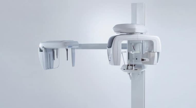
The Morita 3D R100 is exceptional for dental implant planning with full arch 3D imaging, detailed clarity, and low dosage to the patient. T
The i-Dixel 2.0 software from Morita offers advanced dental implant planning features to assist Dr. Reshad. This enables Dr. Reshad to highlight the mandibular canal for easier viewing, measuring the distance to the implant and determining its lingual and buccal position.

Panoramic Imaging
AF Automatic Positioning makes patient positioning almost effortless. A light beam sensor automatically positions the R100 CT scanner without requiring the patient to move. The beam sensor measures the distance to the patient’s teeth; then the arm automatically moves into the most optimal position for imaging.

The DDAE, which is short for Digital Direct Auto Exposure is a function within the ct scanner that controls x-ray emissions in real-time depending on the area being examined and produces a wide dynamic range, as well as vivid and clear images.
To assist in creating the best image possible Auto Image Enhancement computes a logarithmic conversion factor to adjust the overall density and to highlight shaded details.
The Partial Panoramic Function is engaged when a full panoramic image is not required, in order to expose the areas within the region of interest by Dr. Reshad.
High-Resolution Images with Dose Reduction Feature
Clarity and Resolution
The Morita 3D R-100 offers the best in high-resolution images. It provides stunning clear images of the periodontal pocket, the alveolar bone, and the periodontal ligament. It is optimized and built with dental implant therapy in mind, from planning to post-operative observation.
Dosage Reduction
Through technological advancement and engineering, a Dose Reduction feature optimizes the intensity of the x-rays which lowers exposure for easily penetrated tissues. Up to 40 percent of the dosage is reduced when compared to the standard mode. By maximizing efficiency, soft tissue, such as the maxillary sinus membrane and skin, appear sharper and vivid than previous ct scanner technology with fewer artifacts.

Short Radiation Exposure Time
The Morita 3D R100 has a radiation exposure time of 7.4 seconds for panoramic and just 9.4 seconds for 3D images, the patient is only subjected to x-ray radiation for a brief period of time.

Source: Morita

3 Reasons tO Get a CT Scan with the J MORITA 3D R100 before Dental Implants
Getting an examination with the highest fidelity imaging technology available isn’t just smart, it’s essential. The typical x-ray machine found in most dental offices don’t provide the detailed, three-dimensional images necessary to plan and perform a precise surgery with the utmost accuracy and certainty of success. Dental implantations are complex and require both skill, experience, talent, and accurate planning.
1. In-depth Assessment of Implant Suitability
One of the most important roles of a preliminary CT scan before receiving dental implants is to rely on a comprehensive assessment of a patient’s existing dental conditions.
Three-dimensional tomography helps map out the tooth targeted for extraction and replacement. It also precisely pinpoints the location and spatial form of other vital structures such as blood vessels, bones, and major arteries. Nerves and arteries can be avoided while bone can be examined for areas of potential weakness.
Locating, analyzing, and assessing each of these critical structures is the difference between a failed operation and a successful one. Every dental implant procedure begins with careful diagnosis and case planning. This is impossible without the accurate, and complete, three-dimensional images provided by a CT scan.
2. Accurate Jaw Bone Analysis
CT scans play a particularly critical role in determining whether a patient has adequate jaw bone depth to sustain an implant operation. The jaw bone is a three-dimensional structure with diverse areas of depth, width, and length. Simple two-dimensional radiographic techniques, such as x-rays, don’t provide a clear picture of the nuances of each person’s unique jaw structure. In some instances, patient x-ray results can indicate adequate underlying jaw bone structures while in reality, the bone is quite thin.
Likewise, in some cases, an initial x-ray may incorrectly cause a patient to be rejected for conventional dental implants. For accurately measuring bone depth, a CT scan is the best decision.
3. Precision Placement of Dental Implants
Precise placement of dental implants would be significantly complicated without the detailed, three-dimensional images provided by a CT scan.
An implant that is placed incorrectly by even the smallest of margins can result in pain and discomfort, bite imbalances, dental wear, and eventually, complete failure of the implant prosthesis. An implant that accidentally severs a nerve or damages an artery during insertion can result in unnecessary complications. Like any medical procedure, precision is critical. A CT scan can help a prosthodontist and oral surgeon establish the exact location for a dental implant well before surgery begins.
CT Scans Are the Highest Standard of Care
Dentists, Prosthodontists, and other dental professionals are obligated by oath and by education to provide the best possible care to their patients. With computer-guided tomography technology, conventional dental implantation routines, as well as other dental interventions, can be more comfortable, safer, and much more routine.
CT scans like the Morita 3D R100 permanently remove the guesswork involved with implantation procedures by revealing all of the underlying oral and dental structures.
Prosthodontists can plan out with high accuracy not only the location and position of the dental implants themselves, but also how the implants will be inserted to minimize patient pain and discomfort, all the while avoiding blood vessels, nerves, and other fragile parts of the mouth. For patients seeking the best and highest standard of care, CT scans are recommended.